Making the right decisions about how to deal with a lame animal depends on how accurately the cause of the lameness can be diagnosed and localized. It’s important for those caring for these cattle to determine whether a specific lameness case can be dealt with on the farm or needs veterinary treatment.
Lameness diagnosis consists of two parts: 1) Localizing the lameness to a specific limb or structure (such as a joint or hoof), and 2) Determining the root cause (for example, infection or injury). For instance, a swollen joint in a lame cow may be easy to visualize, but whether it is due to an infection or to trauma may not be easily determined. Careful restraint of the animal and examination of the joint for warmth or even dislocations or fractures may help answer some of those questions. In cases where the lameness can be localized to a limb but not easily determined to arise from a specific joint, restraint and palpation of the joints of the limb, moving from top to bottom is useful. Comparing the size of a suspected swollen joint with the corresponding joint on the opposite, unaffected limb is also helpful.
Inspecting for Disease
Lameness arising from the foot presents some unique diagnostic challenges. Swelling immediately above the hooves is often due to footrot if the swelling is symmetrical in nature. If the swelling is over one toe more than the other, it is often an indication of infection in one toe or in the deeper tissues of the foot. Foot rot is also characterized by a foul-smelling, pus-producing wound between the toes. If the examiner is cautious, this can be detected by running a finger between the toes of an animal restrained in a chute. Hairy heel warts are characteristic in their appearance between the toes in the back or sometimes the front of the foot, along with a severe painful reaction when the lesions are touched by the examiner. It’s very important to differentiate between treatable conditions, such as these, and deeper injuries and infection, for which treatment is more involved or even unnecessary.
Examining the Hooves
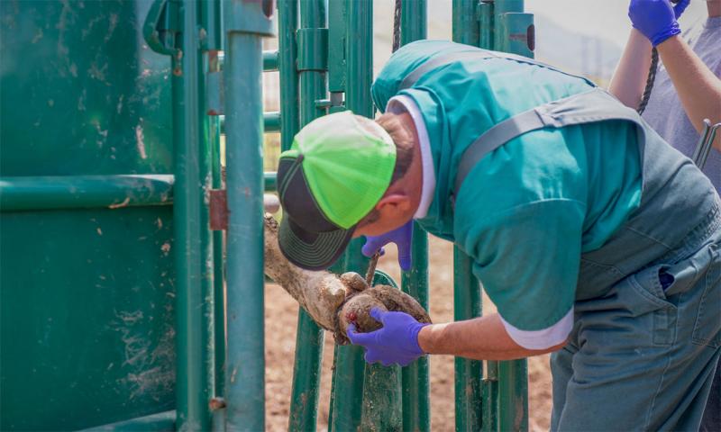
Pinpointing causes of lameness that arise from the foot depends on localizing the pain response within one toe or the other, or sometimes both. This will necessitate examination of the bottom of the hooves. Most cattle chutes used in beef cattle working facilities are not set up for convenient hoof examinations. Yet, failure to examine the bottom of the hoof is a very common way to miss some easily treated conditions (for example, a nail in the foot). In some cases, ropes or straps can be used to lift and immobilize a leg for examination in the chute. Operators should take precautions to avoid injury to themselves and the animal when accomplishing this; beef cattle will tend to vigorously resist such restraint at first, and further injury may be likely.
When there’s any question whether diagnosis or treatment may be beyond the ability of the caretaker, it is preferable to transport cows, bulls and larger calves to a veterinary facility that uses a tilt table. The extra time and expense are invariably well-justified in terms of safety to people and animals. In addition, proper restraint and the right equipment usually results in a much more thorough diagnosis and better treatment outcomes.
Once the bottom of the foot is accessible, it should be cleared of mud and manure with a hoof pick or hoof knife. The bottom of the hoof can be superficially scraped with a hoof knife to better visualize any defects, such as abscesses or penetrating foreign objects.
Locating the Pain Source
If no obvious defects are initially observed, it may be useful to determine which toe is the source of the pain and lameness. Tapping on the bottom of the hoof with a hammer and observing a pain response can help localize the lameness to one toe or the other. Hoof testers, such as those utilized for horses, can also be used. These are pliers-like devices that apply pressure to a localized part of the hoof when squeezed. When pain is not present in the hoof, the animal will not react to the hoof tester. If the tester is squeezed over the painful area, the animal will react by attempting to withdraw the foot or vocalize. If a sole or toe abscess is present, the examiner may even observe pus oozing from the defect in the sole.
Treatment Considerations
Once a cause of lameness is identified, treatment options can be considered. Proper restraint, equipment and expertise are all necessary components if a successful outcome is to occur.